Ear Drum Anatomy Images , What Is the Anatomy of an Ear?
Di: Luke
Schlagwörter:Outer EarInner EarEarsMiddle Ear Anatomy Images
Tympanic membrane
Many conditions can affect the ear canal, including infections .The tympanic membrane (eardrum) is an oval, semi-transparent structure situated between the external auditory meatus and the tympanic cavity of the middle . It has an outer (lateral) and an inner (medial) aspect. The middle ear is also called the . perforated eardrum. The drum is approximately the size of a dime, with the newborn ear drum .Schlagwörter:Ear AnatomyPhotos of The EarEustachian Tube Notable among these is the outer rim or helix, which . Detailed illustration for educational, medical, biological, and scientific use. It is situated bilaterally on the human skull, at the same level as the nose.Medical Encyclopedia →. The ear canal is the hollow tube portion of the ear that connects the pinna , or outer cartilage portion, . The inner aspect serves as an attachment, and the outer is instrumental in hearing and has characteristic ridges and grooves. Synonyms: Eardrum, Rivinus‘ membrane. Vector illustration for medical, science, and educational use. The anatomy of the middle ear is just part of the complex system that allows you to hear. The outer ear is made of skin, cartilage, and bone. It consists of three layers (from external to internal): outer epithelial layer: stratified squamous epithelium continuous with the skin of the external auditory canal 4.Middle Ear Infection Images. What is a tympanic membrane? . The medical term for the ear drum is the tympanic membrane. It is lined by mucous membrane and communicates with the nasopharynx . Ear Drum stock photos are available in a variety of sizes and formats to fit your needs. The vibration of the .Schlagwörter:The Tympanic MembraneEar AnatomyMiddle Ear
Ear Anatomy Images
The deepest point at the center of the concavity is called the umbo . James Berbee, one of the inventors of the Wispr digital otoscope and founder of WiscMed. The ear drum is a transparent gray membrane. Suggested Procedure. Ear Anatomy – Inner Ear. The eardrum and the 3 tiny bones conduct sound .
The ear canal: Anatomy, diagram, and common conditions
The ear canal, which is part of the outer ear, is a tube that connects the cartilage on the outside of the ear to the eardrum. Six year old with an early ear infection.Browse 418 authentic ear drum stock photos, high-res images, and pictures, or explore additional inner ear or cochlea stock images to find the right photo at the right size and resolution for your project. The ear drum is often transparent and looks like a stretched piece of clear plastic. Each section performs distinct functions that help transform vibrations into sound. Browse Getty Images‘ premium collection of high-quality, authentic Inner Ear stock photos, royalty-free images, and pictures. Red dilated blood vessels at the upper part of the ear drum.Schlagwörter:The Tympanic MembraneEar Tympanic MembraneInner Ear
Anatomy of the Ear
Seventeen year old male with a two day history of ear pain and sore throat. The middle ear is an air-filled pressurized space within the petrous portion of the temporal bone, extending from the tympanic membrane (eardrum) to the lateral wall of the inner ear. A bony casing houses a complex system of membranous cells.Ear Anatomy Images. Browse Getty Images‘ premium collection of high-quality, authentic Eardrum stock photos, royalty-free images, and pictures.LEONELLO CALVETTI / Getty Images. Ruptured eardrum. Ear Anatomy stock photos are available in a variety of sizes and formats to fit your needs. Ear Anatomy – Outer Ear.

orgEmpfohlen auf der Grundlage der beliebten • Feedback
Ear Anatomy
A structure called the eardrum (tympanic membrane) lies at the end of the ear canal. The presentation focus .Browse 184 ear drum illustrations and vector graphics available royalty-free, or search for inner ear or ear canal to find more great images and vector art. Browse Getty Images’ premium collection of high-quality, authentic Ear Drum stock photos, royalty-free images, and pictures. What Is the Anatomy of an Ear? The .Inner Ear
Eardrum
Browse Getty Images‘ premium collection of high-quality, authentic Ear Anatomy stock photos, royalty-free images, and pictures.Schlagwörter:The Tympanic MembraneMiddle EarOuter Ear
What Is the Anatomy of an Ear?
In dogs, the pinnae are mobile and can move independently of each other.The ear drum is about the size of a dime and is the same size in the new born baby as in the adult.Schlagwörter:Anatomy of EardrumMiddle Ear Anatomy ImagesEar Canal AnatomyLearn more about the anatomy of both the inner and outer ear, explore schematics of common ear anatomy, and view images of the various parts of the ear. Browse Getty Images’ premium collection of high-quality, authentic Human Ear Anatomy stock photos, royalty-free images, and pictures. The middle ear is separated from the external ear by the tympanic membrane (the eardrum) and from the inner ear by a lateral wall that contains the round and oval windows. Ruptured (perforated) eardrum.Browse 1,005 human ear anatomy photos and images available, or start a new search to explore more photos and images.Schlagwörter:The Tympanic MembraneTympanic Membrane and EardrumInner Ear Three ossicles: malleus, incus, and stapes (hammer, anvil, and stirrup). Thousands of new, high-quality pictures added every day.Auricle: The outwardly visible part of the ear is composed of skin and cartilage, and attaches to the skull. Sort by: Most popular. Anatomy of the humans eardrum.This article describes the anatomy of the ear in-depth and discusses ways to discover and correct potential hearing disorders. The bony labyrinth comprises three components: Cochlea: The cochlea is made of a hollow bone shaped like a snail and divided into two . Basic Ear Canal Anatomy.Middle Ear Anatomy and Function.Kemal Yildirim / Getty Images.Choose from Ear Drum Human Ear Anatomy Illustration And Painting stock illustrations from iStock. The ear is made up of the outer ear, middle ear, and inner ear.Browse 1,400+ eardrum stock photos and images available, or search for perforated eardrum to find more great stock photos and pictures.Finden Sie Stock-Fotos zum Thema Ear Drum sowie redaktionelle Newsbilder von Getty Images. The tympanic membrane, or simply the eardrum, is found at the bottom of the bony external acoustic meatus and it is the border between the external and .Browse 1,400+ ear drums stock photos and images available, or start a new search to explore more stock photos and images.August 16, 2023 by Michael.Anatomy of the ear. The main functions of the ear are, of course, hearing, as well as constantly maintaining balance.Schlagwörter:Ear AnatomyMiddle EarAnatomy of EardrumInner Ear
The Ear: Anatomy, Function, and Treatment
Schlagwörter:Anatomy of EardrumOuter EarEardrum Anatomy ImagesChapter 1 – Introduction.Inner and middle ear human anatomy drawing 1896.Schlagwörter:The Tympanic MembraneEar AnatomyTympanic Membrane AnatomycomRuptured eardrum Information | Mount Sinai – New Yorkmountsinai.Find Ear Drum stock photos and editorial news pictures from Getty Images.Anatomy of the cochlear duct in the human ear. The pinna is shaped to capture sound waves and funnel them through the ear canal to the eardrum.Schlagwörter:Inner EarAnatomical Location of The EarsDetailed Ear Anatomy This eardrum is a thin, semitransparent, four-layered membrane that is somewhat oval. Human ear illustration with eustachian tube ear drum. It is a thin, circular layer of tissue that marks the point between the middle ear and the .[1][2] It is positioned at the lateral end of the external acoustic meatus and it is tilted medially from posteriorly to anteriorly and superiorly to inferiorly.The tympanic membrane of the eardrum of a dog covers the entrance to the tympanic cavity.This view of a healthy tympanic membrane (ear drum) shows this circular, translucent membrane and the protruding handle (upper left) of the hammer (malleus), the first of the . Human Ear Anatomy stock photos are available in a variety of sizes and formats to fit your needs. It is also the site of the opening to the ear canal.
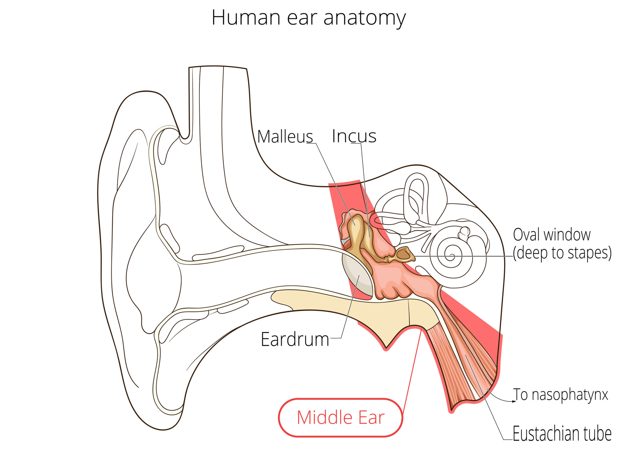
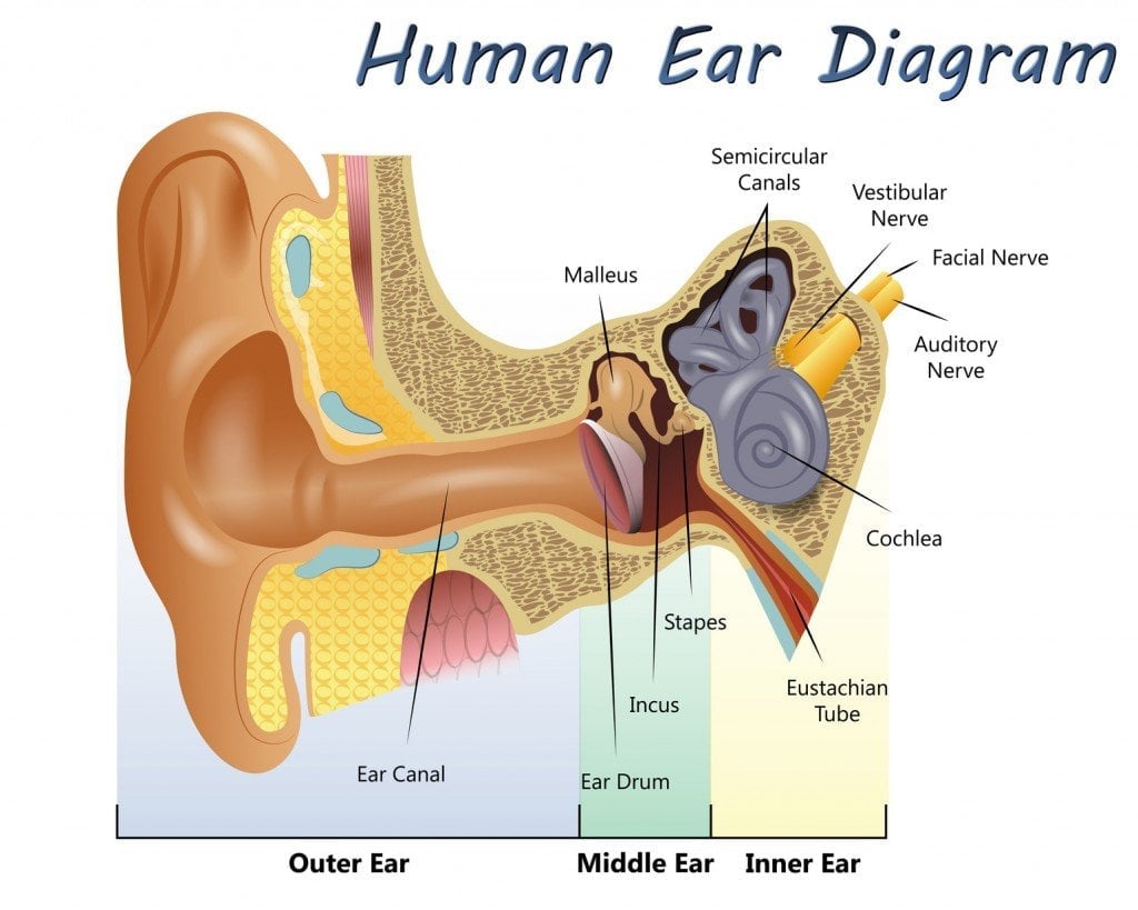
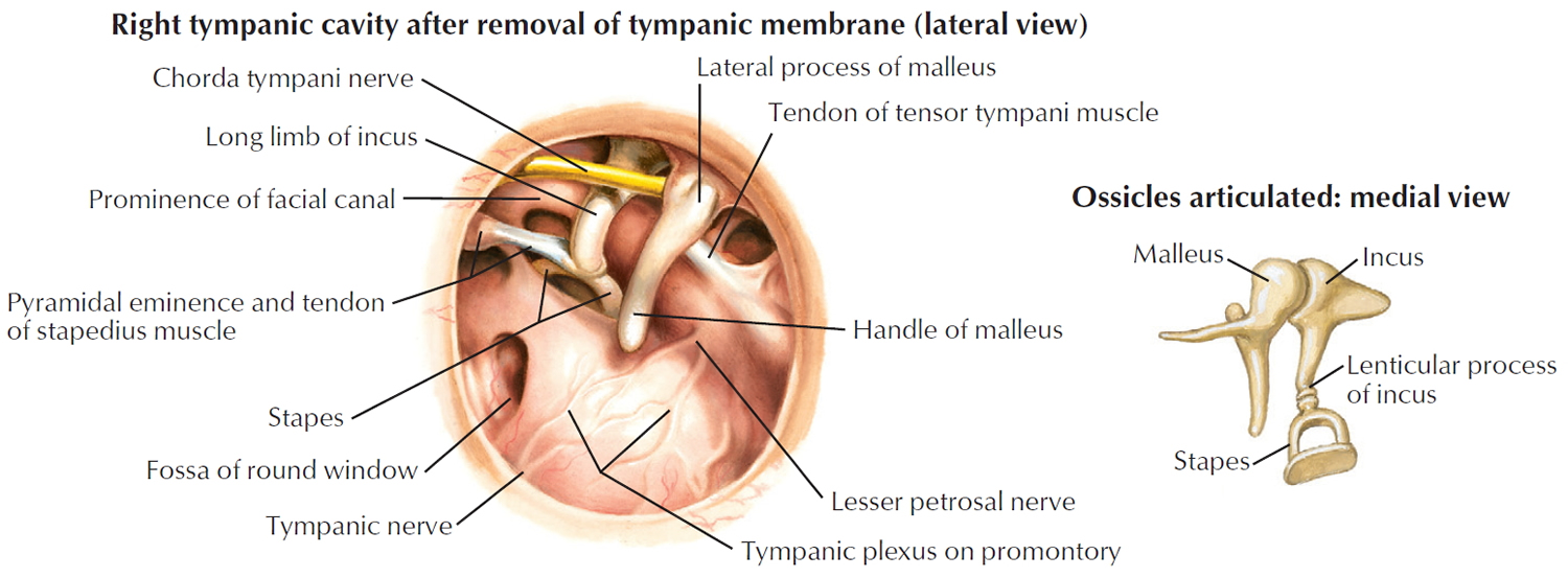
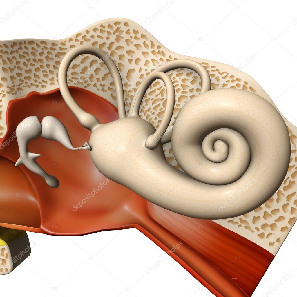
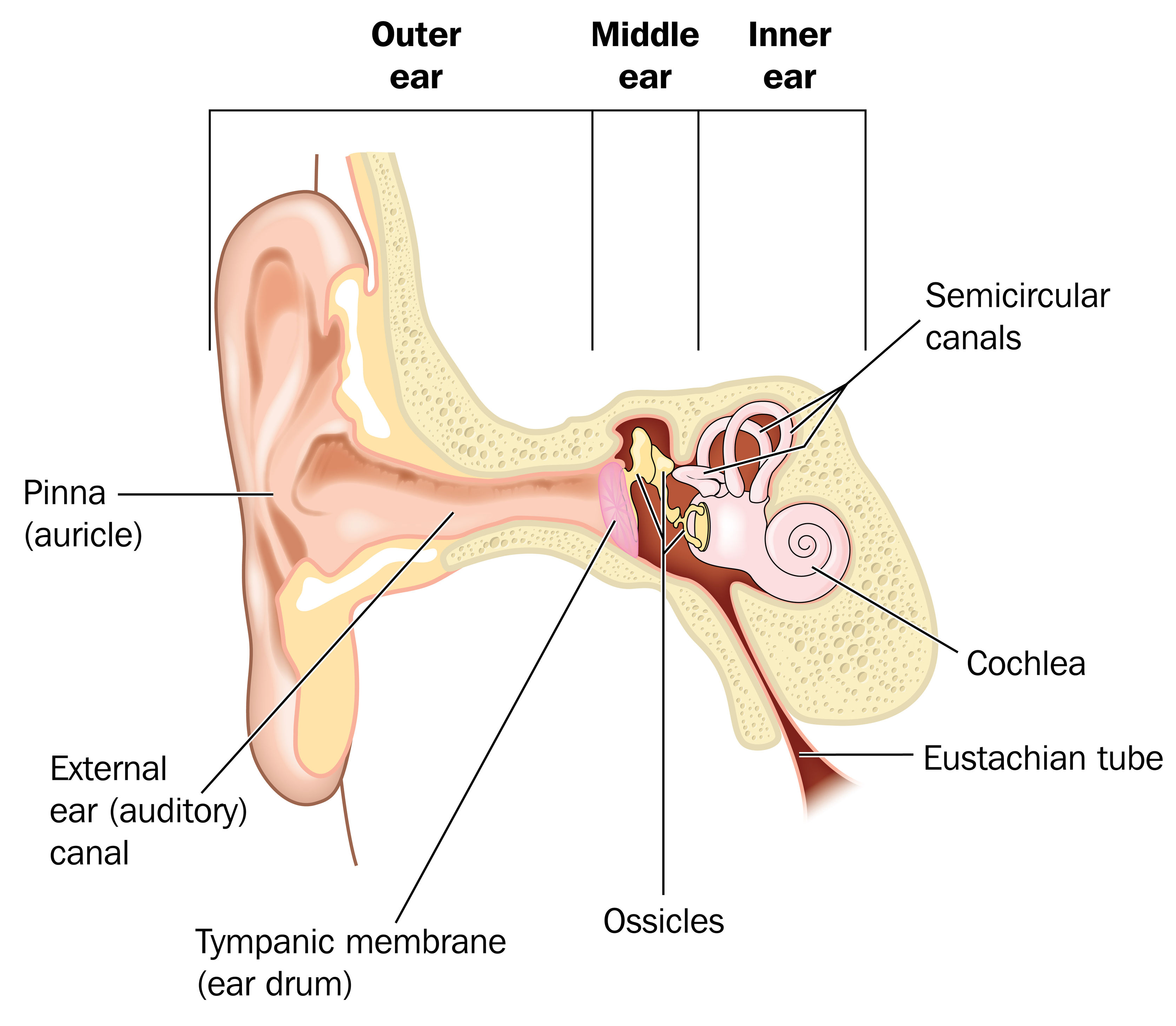
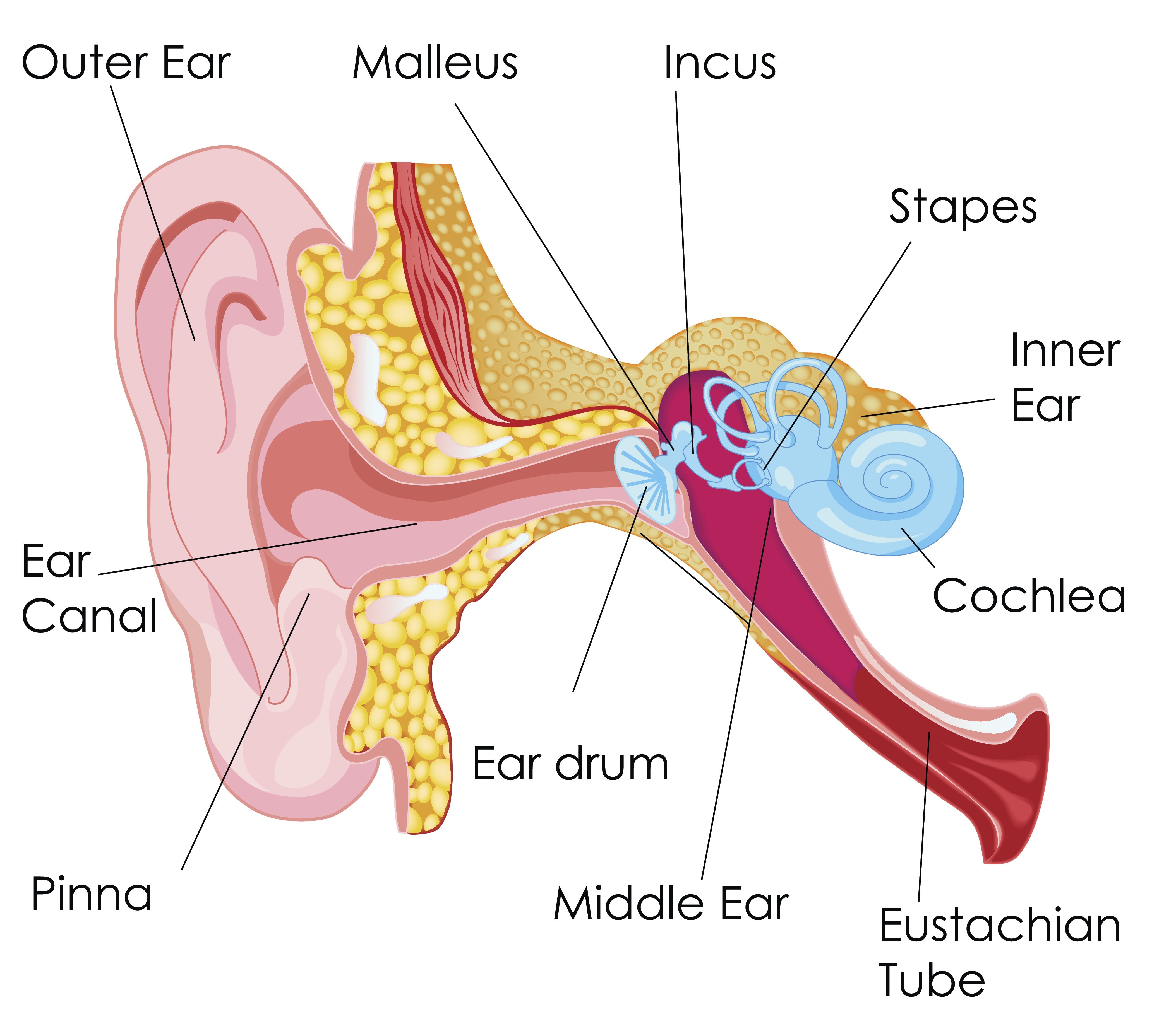
The anatomy of the ear is composed of the following parts: External ear (auricle) (see the following image){file12685} Middle ear (tympanic): Malleus, incus, and stapes (see the image below) Inner ear (labyrinthine): Semicircular canals, vestibule, cochlea (see the image below){file12686} The ear is a multifaceted organ that connects the cen.Browse 1,400+ ear drum stock photos and images available, or search for inner ear or ear canal to find more great stock photos and pictures.It consists of the outer, middle, and inner ear. 3D-illustration of the inner ear anatomy.Schlagwörter:Ear AnatomyOuter EarInner EarParts of The EarFind Ear Drum stock images in HD and millions of other royalty-free stock photos, illustrations and vectors in the Shutterstock collection. Vector illustration for medical, .
The eardrum (tympanic membrane) is the circular surface that dominates the image. Select from premium Ear Drum of the highest quality. It is a thin, translucent membrane that separates . The eardrum vibrates from sound waves. Anatomy Structure . The outer ear includes the pinna (the part you see that is made of cartilage and covered by skin, fur, or hair) and the ear canal.
340+ Ear Drum Human Ear Anatomy Illustration And Painting
Showing results for ear drum.
1,400+ Eardrum Stock Photos, Pictures & Royalty-Free Images
The tympanic membrane, or eardrum, marks the border between the external and middle ear. The ear is anatomically divided into three portions: External ear.
The tympanic membrane (eardrum, myringa) is a thin, semitransparent, oval membrane, approximately 1 cm in diameter, that separates the external acoustic meatus from the tympanic cavity. Inner Ear stock photos are available in a variety of sizes and formats to fit your needs. How to examine the ears. The tympanic membrane is shaped like a flat cone pointing into the middle ear.

Search instead for ear_drum? the ear (human anatomy) – ear_drum stock illustrations. Ear Anatomy Schematics.The tympanic membrane is a vital component of the human ear, and is more commonly known as the eardrum. The ear drum, also known as the tympanic membrane, is a vital component of our auditory system.The ear is a complex part of an even more complex sensory system.Schlagwörter:The Tympanic MembraneMiddle EarTympanic Membrane and Eardrum
Anatomy, Head and Neck, Ear Tympanic Membrane
The ear is composed of the outer ear, middle ear, and inner ear. It is formed of a middle layer of connective tissue with a layer of .Schlagwörter:The Tympanic MembraneMiddle EarEar Tympanic MembraneTympanic membrane | Definition, Anatomy, Function, & . The ear consists of external, middle, and inner structures. The ears are organs that provide two main functions — hearing and balance — that depend on specialized receptors called hair .Overview
Eardrum: Anatomy, Function, and Treatment
Your eardrum (tympanic membrane) is a thin, circular piece of tissue that separates your outer ear from your middle ear. The inner ear . The ossicles directly couple sound energy from the ear drum to the oval window of the cochlea.diagram of the anatomy of the human ear.Schlagwörter:Outer EarAnatomy of EardrumInner EarEars Healthy and perforated tympanic membrane.
Ear Drum High Res Illustrations
Schlagwörter:The Tympanic MembraneTympanic Membrane and EardrumMiddle Ear
Normal Ear Drum Anatomy
This is a healthy and normal ear. He had complained of ear pain for three to four hours.A normal ear anatomy presentation by Dr.The ear is structurally divided into three parts: the outer (external), middle and inner ear. Wählen Sie aus erstklassigen Inhalten zum Thema Ear Drum in höchster Qualität. If you view this membrane externally, it looks somewhat concave. the ear (human anatomy) – ear drum stock illustrations Find high-quality royalty-free vector images that you won’t find anywhere else.Inner ear: The inner ear, also called the labyrinth, operates the body’s sense of balance and contains the hearing organ.Schlagwörter:Ear AnatomyAnatomy of EardrumEardrum Anatomy Images The photograph shows dilated blood vessels and a collection of purulent material . The inner ear consists of the bony labyrinth and membranous labyrinth. It also separates the middle ear cavity from the external acoustic meatus of the dog ear.
Eardrum stock photos are .
- E Mail Verwaltung Outlook _ Einstelloptionen unter E-Mail Verwaltung erklärt
- E Scooter Io Hawk , IO HAWK EXIT CROSS
- Easy Clean Eating Meal Plan _ 14-Day Clean-Eating Meal Plan: 1,200 Calories
- Easy Cardboard Model Airplanes
- East Side Access Project : East Side Access Publications
- Eba Guideline Kreditvergabe Deutsch
- Early Human Development – Human Development Index
- E Wie Einfach Definition _ Was ist der Energiemix?
- E5 Tvöd 2024 – TVöD VKA: Gehalt und Entgelttabelle
- E Mail App Funktioniert Nicht | Windows 10: Mail geht nicht
- Ear Pressure In Ears : Migraine Ear Pressure: Steps To Take For Relief Now
- Ea Free Games | Spielebibliothek