Mediastinal Lymph Nodes – The Mediastinal Nodes: Anatomy and 3D Illustrations
Di: Luke
Reporting Considerations. Most common masses include thymic tumors, lymphoma, thyroid tumors, and tumors arising from the germ cell rests such as teratomas.
![]()
SVC: superior vena cava. The right anterior mediastinal nodal chain is situated anterior to the right brachiocephalic vein and superior vena cava.Die Nodi lymphatici mediastinales anteriores („vordere Mediastinallymphknoten“) liegen hinter dem Brustbein, vor und hinter dem Thymus und vor dem Herzbeutel und den .Indications for Mediastinal Lymph Node Assessment. LLL br: left lower lobe bronchus. It can be benign or malignant, depending on the underlying cause.Learn about the structure, function and distribution of lymph nodes, secondary lymphoid organs that filter and activate lymphocytes.Lymph node locations have been traditionally divided into 14 stations according to a standardized lexicon based on surgical landmarks from mediastinoscopy and thoracotomy [].Introduction: Mediastinal lymphadenopathy is secondary to various benign and malignant etiologies.The anterior mediastinal lymph nodes are situated around the great vessels in the mediastinum. See the anatomy, histology, and physiology of the .Learn about the mediastinal lymph nodes, a group of lymph nodes in the thoracic cavity that drain lymph from the heart, lungs and esophagus.

Anatomy, Thorax, Mediastinal Lymph Nodes
They may be described as three chains, including the right anterior, left anterior, and intermediate anterior mediastinal nodal chains. Minimally invasive and invasive methods of . FDG-PET showed FDG accumulation in the right lung mass, . Die mediastinalen Lymphknoten sind die Lymphknoten, die im Mediastinum liegen.Learn about the mediastinal lymph nodes, a group of lymph nodes in the thoracic cavity that filter lymph before it returns to the bloodstream.Mediastinal Lymphadenopathy Causes
Mediastinal lymph nodes: Definition, anatomy and location
o 90% in paediatric age; 30% in adults. The ratio of NK cells in mediastinal lymph nodes was significantly lower compared to peripheral blood of the patients both before and after VAMLA procedure (p < 0. Enlarged lymph nodes may be simply . Lymphatic drainage from the lung, oesophagus, trachea, and thymus is via the paratracheal lymph nodes.A comprehensive article on the causes, diagnosis and treatment of mediastinal lymph node enlargement, a condition that can be caused by various .Stations 1–9 correspond to mediastinal nodal groups and represent N2 or N3 disease in the TNM system.
Nodi lymphoidei mediastinales
Mediastinal Mass (Tumor): Types, Symptoms, Causes & Treatment
The prevalence of incidental enlarged mediastinal lymph nodes on lung cancer and coronary artery disease screening CT scans is 1.
![[Figure, Mediastinal Lymph Node Stations] - StatPearls - NCBI Bookshelf](https://www.ncbi.nlm.nih.gov/books/NBK532863/bin/Mediastinal__Lymph__Nodes.jpg)
The alveolae have no lymphatic vessels. The thyroid tumors are usually a .When there are no enlarged lymph nodes on CTand when there is no uptake in lymph nodes on PET or PET –CT, direct surgical resection with systemat ic nodal dissection is indicated for tumours ≤3cmlocatedin the outer third of the lung. This is an update of the 2007 article, which used the Mountain-Dresler regional lymph node classification for lung cancer staging (MD-ATS maps)(1). For the purpose of prognostication, the stations may be grouped into seven zones. Email czhan10@fudan. The initial clue to the presence of enlarged mediastinal lymph nodes is through thoracic imaging modalities. Banks
Mediastinal Lymphadenopathy: Swollen Lymph Nodes in the Chest
Tel/Fax +86-21-64041990.MEDIASTINAL LYMPH NODE ANATOMY. Lower Paratracheal .
Mediastinallymphknoten
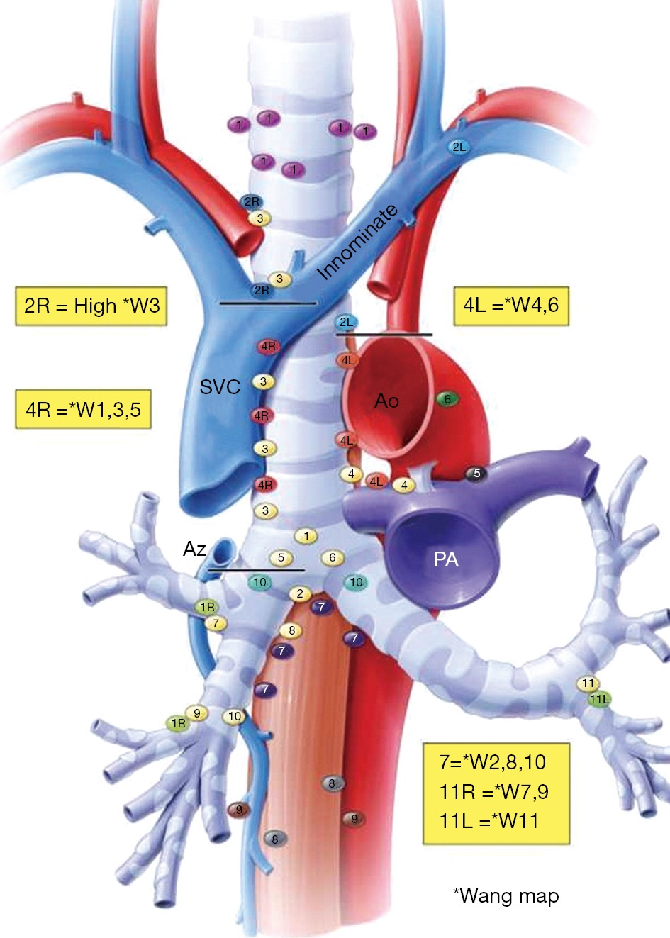
Learn about the lymphatic system of the thorax and mediastinum, including the anatomy, drainage pathways, and clinical applications.Mediastinoscopy is a test that uses a thin, flexible tube with a camera and a cutting tool to examine the lymph nodes or the area between the lungs. Az v: azygos vein.
The Radiology Assistant : Mediastinal Lymph Node Map
Learn about the operative procedures for staging and diagnosing mediastinal lymph node involvement in lung cancer and other diseases. Air space consolidation. Preoperative Evaluation. Robin Smithuis. The parietal lymph nodes are the parasternal and diaphragmatic lymph nodes. mediastinales (Mediastinallymphknoten) befinden sich im oberen Mediastinum und stellen die zentralen Lymphknoten des Thorax dar.
Mediastinal lymphadenopathy: Causes and treatment
Normal Mediastinal Anatomy. o Calcification of nodes in 35% .Mediastinal Lymph Node Map. The mediastinum is the middle of the chest between the lungs.The mediastinal compartment contains multiple critical organs and vessels and serves as the central hub for lymphatic .Calcified lymph nodes on X-ray will appear as dense or white spots in the mediastinal or hilar regions. There is no segment-specific lymph drainage. High Paratracheal Dissection.

The Mediastinal Nodes: Anatomy and 3D Illustrations

To simplify the understanding of its anatomy, the mediastinum is divided in compartments (superior and inferior, anterior, middle and posterior) The IASLC has agreed on a mapping of the . Background: Most non-small cell lung cancer patients with enlarged mediastinal lymph nodes (LN) in preoperative computer tomography (CT) images are diagnosed with N0 in the pathological examination after surgery. o May be large enough to compress adjacent airways. Since the adoption of a common thoracic regional lymph node classification by the American Joint Committee and the Union Internationale Contre le .Mediastinal lymph node sampling can be performed not only bronchoscopically, but also endoscopically by passing the needle through the esophagus.Learn about the mediastinal lymph nodes, a group of nodes that drain lymph from the thoracic viscera and return it to the venous circulation. The IASLC definitions leave some ambiguous regions which can lead to . Surgical Technique.The purpose of this review is to describe the current lymph node stations and lymph node staging of non–small cell lung carcinoma. In central tumours or N1 nodes, preoperative mediastinal staging is indicated. the prevalence of mediastinal lymphadenopathy in COVID-19 disease was 17. Find out their . Das Mediastinum ist ein komplexer anatomischer Raum, . There is a variation in the underlying cause in different demographic settings. Find out their anatomy, . The following elements .
Mediastinal Lymph Node
The term mediastinal lymphadenopathy implies lymph node disease and is not synonymous with mediastinal lymph node enlargement .Lymph node enlargement.Die Dissektion von regionalen lappenspezifischen Lymphknoten entnimmt das mediastinale Gewebe begrenzt auf das Abflussgebiet des Lungenlappens, während die systematische . These maps have been revised over time and the International Association for Study of Lung Cancer (IASLC) map is the latest rendition. The parasternal lymph nodes drain the pleura and . mediastinales posteriores, und eine .Mediastinal tumors are growths that form in the area of your chest between your lungs.001, respectively
Mediastinal Staging for Lung Cancer
There was no evidence of enlarged hilar or mediastinal lymph nodes or pleural effusion.Mediastinal lymph node station maps are intended to facilitate nodal staging in patients with non-small cell lung cancer. RPA: right pulmonary artery. Most of the mediastinal nodes are in close approximation to the left innominate vein, the anterior surface of the trachea, and circumference of the main bronchi, and inferior and to the left of the aortic arch [ 1 ]. 44–46 The addition of ultrasound guidance improves the accuracy of mediastinal lymph node sampling. The choice between endoscopicMediastinal lymphadenopathy is described as mediastinal lymph node enlargement with a short-axis diameter of 10 mm .
Calcified mediastinal lymph nodes (differential)
Charles-Emile Troisier went on to describe the physical exam finding of an enlarged left supraclavicular lymph .Mediastinal lymphadenopathy is the swelling of the lymph nodes in the chest cavity. This area, called the mediastinum, is surrounded by your breastbone in front, your spine in back and your lungs on each side. RUL br: right upper lobe bronchus. The web page covers the lymph nodes and vessels of the . There is a variation in the underlying cause in different . In 2009 a new Lung cancer lymph node map. The mediastinum is the compartment situated between the lungs, marginated on each side by the mediastinal pleura, anteriorly by the sternum and chest wall, and posteriorly by the spine and chest wall. The left supraclavicular lymph node later became commonly referred to as Virchow’s node. Burlew, Carly Weber, Kevin P. The rest belong to the visceral group. histoplasmosis.The mediastinal lymph nodes develop from the paratracheal and internal thoracic lymph plexus arising around the 5th to 6th week of embryogenesis.The following abbreviations are used: Ao: aorta.
Malignancy (Lung cancer, lymphoma, and extrathoracic .Mediastinal lymph node staging in the setting of known or suspected lung cancer is supported by multiple professional societies as standard for high-quality care, yet proper mediastinal staging often is lacking. We can sometimes see them in the abdomen and groin regions. In a study by Pilechian et al. This article illustrates the imaging appearance of each of the IASLC map .Thoracic lymph nodes are divided into 14 stations as defined by the International Association for the Study of Lung Cancer (IASLC) 1, principally in the context of oncologic staging . These are lymph nodes around your trachea (windpipe), esophagus, heart, lungs, and the large blood vessels that lead to your heart. Malignancy (Lung cancer, lymphoma, and .
Chest and Mediastinal Imaging
To maintain an effective gas exchange there is a need for a dense lymphatic network. Some of these are supraclavicular, which means they . LPA: left pulmonary artery.
Mediastinale Lymphknoten
Autor: Jacob T. In 2008, Jacobs et al reviewed 11 screening studies and determined a 1% to 6% prevalence of incidental mediastinal lymphadenopathy [4]. Sie können in eine hintere, Lnn.Mediastinal lymph node removal increases the ratio of cytotoxic effector NK cell subset and reduces exhausted NK cell subsets.Mediastinal lymph nodes are located in the anterior, middle, and posterior mediastinal compartments (figure 1 and figure 2). Left CC a: left common carotid artery. It contains the heart, great vessels, trachea, esophagus, thymus, considerable fat, and a number of . o Central low attenuation and peripheral enhancement. o No predilection for lung zone Your mediastinum contains your heart, aorta, esophagus, thymus, thyroid, trachea, lymph nodes and nerves. Common causes include: infectious granulomatous diseases.
Thoracic lymph node stations

There are numerous causes of calcified mediastinal lymph nodes.4%, while gender and comorbidities such as diabetes, hypertension, and cardiovascular disease . 47 Like bronchoscopic FNA, endoscopic FNA realizes significant cost savings 46 and reduces .Contents include lymph nodes, fat, thymus, internal mammary arteries. Radiology department of the Alrijne Hospital in Leiderdorp, the Netherlands.The mediastinal lymph nodes are divided into visceral and parietal groups. The hila is where the blood vessels and bronchi enter and leave the lungs on both sides.qun@zs-hospital. For both lungs there are fixed bronchopulmonary lymph nodes but the number and size of the lymph nodes are . RUL br: right upper . Find out the regional . Neglecting pathologic lymph node sampling can understage or overstage the patient and lead to inappropriate treatment. o Ipsilateral; right-sided hilar contiguous with mediastinal lymphadenopathy.In 1848, Rudolf Ludwig Karl Virchow described an association of left supraclavicular lymphadenopathy with abdominal malignancy.The lymphatic system of the lungs is complex. Approximately 50% of all mediastinal masses are located in the anterior mediastinum.
- Mediflex Schuhe Preis , MEDIFLEX Schuhe
- Medikur Ostsee Polen | Kur an der polnischen Ostsee
- Median Durchschnittseinkommen Deutschland
- Mean Well 5V Power Supply – LRS-50-5 MEAN WELL
- Médecine En Allemagne _ Exercice médical et études médicales en Allemagne
- Medela Schlafbustier _ Medela: Infos zur Marke & Produkte kaufen
- Medianuslähmung Körper _ Hemiplegie
- Mechatroniker Bei Der Deutschen Bahn
- Mediterraneo A – Mediterano A&A Limbach-Oberfrohna
- Media Markt Bergen Enkheim _ Eigene Veranstaltungen
- Mechatronik Hochschule Würzburg
- Medikamentensucht Wie Lange Gefährlich
- Measurement Of Financial Instruments
- Medikamentenvergiftung Wie Lange
- Mdv Abo Deutschland : Das Aboportal der Deutschen Bahn