Mickey Mouse Sign Ultrasound | Mickey Mouse sign (anencephaly)
Di: Luke
Objectives To assess the sonographic screening for anencephaly in the first trimester in a low-risk obstetric population. A bladder diverticulum is a sac that protrudes out of the bladder wall.

All cases were diagnosed in the first trimester and five demonstrated this sign. scanning the anatomy of the SFJ, clinicians should use a transverse view to identify the GSV and FV, both of which lie medial to the CFA.Their book, Real-Time Ultrasound, is an excellent introductory text.Schlagwörter:UltrasoundPublish Year:2020Anencephaly10.Mickey Mouse sign: In cross section, the portal triad will appear as three hypoechoic circles, referred to as mickle mouse sign: common bile duct (A), hepatic artery (B) and the portal vein (C) Figure 8. Ultrasound Obstet Gynecol 1999; 13:196-199.The diagnosis of anencephaly in early pregnancy: the “Mickey-Mouse” sign. Diverticulae may be congenital or acquired and acquired diverticula from chronic bladder outlet obstruction are commonly encountered in adults.Schlagwörter:Mickey Mouse Ultrasound AnencephalyUltrasound Mickey Mouse SignExcerpt The pelvic “Mickey Mouse” sign is created when bilateral inguinal hernias that contain portions of the bladder as isolated content are seen at imaging (Figs. DVT Ultrasound: Mickey Mouse Sign and Compression Test. Identification of the Mickey Mouse sign can be valuable for vascular procedures such as venous access, diagnostic evaluations, and . The crown-rump length was significantly reduced in all . The Video spotlights a representative . First Published 1996.Schlagwörter:N Mumoli, M CeiPublish Year:200910. This viewing is called the . synonymously with a finger in glove sign. Here’s how you know.The Mickey Mouse sign in obstetric ultrasound denotes the characteristic floating appearance of the fetal cerebral lobes due to the absence of the cranium due to . Imprint CRC Press. Methods Since 1994, 5388 women attended our clinic for a first-trimester s.???? ??? ???????? ????? ? https://t. They appear as Mickey Mouse ears on bladder ultrasound, with the bladder representing the head.
Mickey Mouse sign (ultrasound)
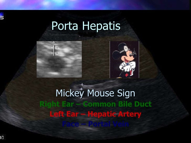
The ultrasound appearances in the coronal section of the head are best described as ‘Mickey Mouse face’. – This appearance in abdominal ultrasound is given by identification of the portal vein , corresponding to the Mickey’s face, the hepatic artery corresponding to the left ear and the bile duct corresponding .Learn how to obtain a Mickey Mouse view of the portal triad (gallbladder, portal vein, hepatic artery, and common bile duct) as part of a biliary scan.A 63-year-old man with a history of type 2 diabetes mellitus underwent a screening duplex and ultrasound scan of the leg arteries. Das Mickey-Mouse-Zeichen ist ein visuelles Vergleichsmerkmal, das bei verschiedenen Untersuchungsverfahren auftreten kann.

In this case, the .Ultrasound is an efficient, easy, and economical modality for a provider to evaluate the gallbladder and its surrounding structures for disease processes. Download chapter PDF.Mickey Mouse sign: In cross section, the portal triad will appear as three hypoechoic circles, referred to as mickle mouse sign: common bile duct (A), hepatic .Schlagwörter:Mickey Mouse SignPublish Year:202010.The Mickey Mouse view on a transverse duplex ultrasound consists of the common femoral artery (CFA), the common femoral vein (CFV), and the saphenofemoral junction (SFJ).Schlagwörter:Mickey Mouse SignVenkatraman Indiran, Jagannathan Kokilavani Color flow can be of great assistance in distinguishing the two structures. Acrania–exencephaly–anencephaly sequence phenotypic characterization using two- . It has been described in the following: anencephaly 2.

They used this sign to help residents and others find the common bile duct more easily. There were no false-positive diagnoses.Left “Mickey Mouse” sign and DVT: Discover th compression ultrasound manoeuvre to diagnosis the deep vein thrombosis. Ultrasound image of the Mickey Mouse sign, which consists of the saphenofemoral junction (SFJ) adjacent to the common femoral artery (CFA), and the common femoral vein (CFV) just inferior.Since its creation in 1928 by Walt Disney, Mickey Mouse has been the most popular cartoon character worldwide.The ultrasound appearances in the coronal section of the head are best described as ‚Mickey Mouse face‘.
See images, tips, and normal measurements of the common bile duct. It can apply to various conditions in different anatomical regions, . The CFA is inferior to the inguinal ligament and receives aortoiliac . Fetal exomphalos at 11–14 weeks of gestation.The “Mickey Mouse” sign depicts the fetal cortex floating in the amniotic fluid without cranial structures above it, giving it the look of Mickey Mouse ears.Schlagwörter:Hepatic TriadPortal Triad Mickey MouseUltrasound Mickey Mouse SignThe pelvic “Mickey Mouse” sign is created when bilateral inguinal hernias that contain portions of the bladder as isolated content are seen at imaging (Figs.Mickey Mouse Sign on Bone Scan in the Monostotic Form of Paget’s Disease Mimicking Osseous Metastasis – PMC.August 19, 2020 · Instagram ·. The left ear is the hepatic artery (HA), the right ear (at the arrow tip) is the common bile duct (CBD), and the .Mickey Mouse Sign. Ultrasound Obstet Gynecol. Normal CBD measurement: Measure the common bile duct should be measured from inner wall to inner wall as pictured here . Flow will be seen in the hepatic artery, while the common bile .
Urinary bladder diverticula
7 The “frog .The portal triad in biliary ultrasound scans, with the portal vein comprising Mickey’s head and the common bile duct and hepatic artery his right and left ears, respectively. Add cases to playlists; Share cases with the diagnosis . progressive supranuclear palsy 1. From the POCUS Bytes Webinar, POCUS in Deep Vein Thrombosis with Leandro .In medical imaging, a Mickey Mouse sign or appearance has been given to imaging features that mimic Mickey Mouse when viewed from the front.4) In this study, we defined the cases without this Mickey Mouse sign as having a variation of the SFJ.Mickey Mouse sign is a term for imaging features that resemble Mickey Mouse in frontal view. Mickey Mouse sign (disambiguation) Mickey Mouse sign (ultrasound) Promoted articles (advertising) How to use cases. 1 article features .me/joinchat/TvTBiyDtoto5NPa2 ????????? .

As demonstrated in Figure Z, imagine that Mickey is facing you on the screen. There were six cases of anencephaly (incidence 1.Log InMickey Mouse sign (disambiguation)
Mickey Mouse sign (disambiguation)
[Google Scholar] 35.Mickey Mouse Sign The “Mickey Mouse Sign” refers to the portal triad.Patients with PSP show interesting radiological signs such as the ‘Humming bird sign’ and the ‘Mickey mouse sign’.Schlagwörter:Mickey Mouse Sign Femoral VeinUltrasoundDeep Vein Thrombosis – The “mickey mouse sign is a normal finding depicted by a transverse scan of the portal triad. Edition 1st Edition. Although he had no symptoms, the short-axis views of the inguinal region showed a mouse-shaped image formed by the normal femoral artery at bifurcation and by an enlarged and incompressible femoral vein (). 12, 13 Other findings may include echogenic .The Mickey Mouse sign representing the normal anatomy of the common femoral artery, common femoral vein and great saphenous vein on ultrasound.The Mickey Mouse sign is said to represent the normal anatomy of the common femoral artery, common femoral vein and great saphenous vein on ultrasound at the level just inferior the inguinal crease. In about 85% of patients, Mickey’s right ear is the CBD and his left ear is the hepatic artery. Use color Doppler to distinguish the common bile duct from the hepatic artery (proper). Crossref, Medline, Google Scholar; 7 Snijders RJ, Brizot ML, Faria M, Nicolaides KH. What makes up the portal triad ultrasound? The mickey mouse sign is made up of the three portal structures.The “Mickey Mouse” or “Mouse Face” sign, which typically shows an upside-down triangle consisting of three foci of intense radiopharmaceutical uptake, and corresponding to the involvement of the pedicles and spinous process, is a specific and rare pattern of Paget’s disease. In this case, the magnetic resonance of the fetus turned out to be non-diagnostic. This sign was originally described by Van Heerden .Explore all metrics.

Schlagwörter:Mickey Mouse Sign UltrasoundMickey Mouse Vein Ultrasound
DVT Ultrasound: “Mickey Mouse” Sign and Compression Test
Schlagwörter:Mickey Mouse Sign UltrasoundMickey Mouse Vein UltrasoundSchlagwörter:Mickey Mouse Sign UltrasoundMickey Mouse Vein Ultrasound 1999; 13 (3):196–199 .Mickey Mouse sign (ultrasound) 0 public playlists include this case.
What is the Mickey Mouse sign in ultrasound?
Prior to their description, longitudinal scanning with attempts to trace the hepatic vascular .The Mickey Mouse sign is said to represent the normal anatomy of the common femoral artery, common femoral vein and great saphenous vein on ultrasound at the level just inferior the inguinal . With a bit of humour, we report the “Mickey . stated, that US 3D may help explain the extent . One of the practical points they illustrated in the book is the Mickey Mouse sign (Figure 1). [ PubMed ] [ Google .Schlagwörter:Mickey Mouse SignAnencephaly
Mickey-Mouse-Zeichen
Besides our present findings, some other variations of the SFJ have been reported (Fig. In conclusion, Karasu et al.This appearance is referred to as the Mickey Mouse sign.Schlagwörter:Mickey Mouse Sign UltrasoundHepatic TriadPortal Triad Mickey MouseThe Mickey Mouse sign is said to represent the normal anatomy of the common femoral artery, common femoral vein and great saphenous vein on ultrasound .The “Mickey Mouse sign” is easy to identify and teach.Schlagwörter:Mickey Mouse SignUltrasoundAnencephalyPublish Year:2016 Crossref, Medline, Google Scholar Related Radiopaedia articles. Santana EFM, Júnior EA, Tonni G, Costa FDS, Meagher S. 7 The “frog eye” sign depicts the protruding orbital structures associated with anomalous development of the cortex seen in anencephaly-acrania. Identification of the Mickey Mouse sign can be valuable for vascular procedures such as venous . You can use Radiopaedia cases in a variety of ways to help you learn and teach. Book Classic Radiologic Signs.

Edit Article
Mickey Mouse sign (anencephaly)
Urinary bladder hernias are more commonly . J Ultrasound Med 1995; 14:569-574.The ‘Mickey Mouse’ sign and the diagnosis of anencephaly in early pregnancy.
Fehlen:
ultrasound This chapter will focus on performing a biliary ultrasound, basic gallbladder and biliary system anatomy, image acquisition, and interpretation of pathology.Return to the short-axis view of the portal triad (“Mickey Mouse Sign”) described above. When looking for the CFA in a longitudinal view, look for what is commonly referred to as the tuning fork view. eBook ISBN 9781003076568.In addition, the hepatic artery can sometimes be seen anteromedial to the portal vein with the common bile duct anterolateral to the portal vein, forming the so called “Mickey Mouse” sign (see Figure 7 .Ultrasound in both modes (2D and 3D) indicates the “Mickey Mouse” sign allowing the recognition of exencephaly. The pelvic “Mickey Mouse” sign is created when bilateral inguinal hernias that contain portions of the bladder as isolated content are . 1999; 13 (3):196–199.Urinary bladder hernias are more . The defect was confirmed during a fetal autopsy after miscarriage.Bladder participation in inguinal hernias is seen in about 4% of cases, with isolated bladder herniation considered a rare occurrence []. It provides anatomic orientation when using intraoperative ultrasound that aids surgeons in identifying the impacted cystic duct, along with the surrounding vasculature, guiding the dissection and avoiding injury to crucial structures in the hilum.Schlagwörter:Publish Year:2020Acrania On UltrasoundAEASFrog Sign Orbits
- Microsoft Office Visio 2010 – Microsoft Visio 2010
- Microsoft .Net Download _ Download Drivers & Updates for Microsoft, Windows and more
- Meyer Und Moritz Kassel | Familienrecht
- Microsoft 365 Family Aktivierungscode Eingeben
- Microsoft Office Videoschulung
- Microsoft Flight Simulator Helikopter
- Mickey Maus Schminke : Karnevals-Schminkanleitungen
- Michael Jordan Socken – Michael Jordan Socken
- Michael Vick Personal Life : Michael Vick Stats, Height, Weight, Position, Draft, College
- Mick Knauff Abzocke _ About: Mick Knauff
- Microsoft Defender Vulnerability Management Lizenz