Proton Nmr Spectra – Proton nuclear magnetic resonance
Di: Luke
Notice the appearance of both free NH NH bands (sharp, 3300 3300 – 3500cm−1 3500 cm .Lots of Additional Resources for Solving NMR Spectra. At minimum, the spectral window should be 1 ppm to 9 ppm – for 1 H NMR and -10 ppm to 180 ppm for 13 C NMR.10 Overlap in Proton NMR Spectra. In the above spectrum, it is the same proton spectrum for propyl acetate on each of the axis because it is a homonuclear experiment where you look at the same nuclei. The computer in an NMR instrument can be instructed to automatically integrate the area under a signal or group of signals. Magic-angle spinning alleviates this broadening by inducing coherent averaging.The 1 H NMR spectrum of dipropyl ether shows three signals with the triplet at 3. Now that we know the details of NMR, let’s make more specific connections. You could refer to the chemical shift table above to decide where the peaks are likely to be found, but it isn’t really necessary. Proton NMR vs carbon NMR. Proton NMR practice 3 . Figure 24-1: Infrared spectra of propanamide, N N -phenylethanamide, and N N, N N -dimethylmethanamide in chloroform solution. Proton NMR practice 2 .Sometimes the N−H N − H resonance has nearly the same chemical shift as the resonances of CH3−C CH 3 − C protons (as with N N -ethylethanamine, Figure 23-5).
Boosting the resolution of multidimensional NMR spectra by
– Solid-state NMR. This means that this carbon does not have .Proton nuclear magnetic resonance spectroscopy (1 H-NMR) is well suited for the determination of explosives because these analytes are small aromatic molecules which . The peaks are displayed on the axis. In a high resolution spectrum, you find that many of what looked like single peaks in the low resolution spectrum are split into clusters of peaks.
Try the new HTML5 only predictor that works also on iPad, Android, . a quartet counts as only one signal).Online Spectral Database: Quick access to millions of NMR, IR, Raman, UV-Vis, and Mass Spectra. Spectra for dilute solutions are quite easy because of the high sensitivity of the hydrogen nucleus.Overview
Simulate and predict NMR spectra
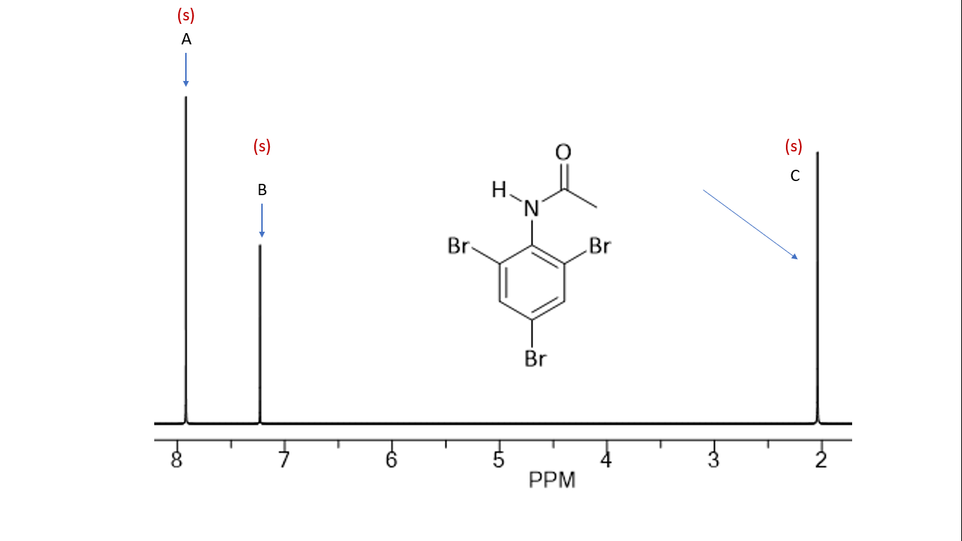
Figure 5: 1 H solution NMR spectrum of acetic acid.Figure 5 provides an example of a proton (1 H) NMR spectrum, meaning that only the protons of the molecule are detected. The signal at 3. The peak at 171 ppm in the 13 C has no cross peak. Carey 4 th Edition On-Line Activity.eduEmpfohlen auf der Grundlage der beliebten • Feedback
How to Interpret Proton NMR Spectra
Unter Attached Proton Test (engl., kurz APT) versteht man ein Kernresonanzspektroskopieverfahren, oder auch NMR-Experiment (Messmethode . and does not require JAVA (only HTML5)!!! This page . On this page we are focussing on the magnetic behaviour of hydrogen nuclei – hence the . For example, in the NMR spectrum of 3,3-dimethyl-1-butyne, the terminal hydrogen of the alkyne appears at ? = 2.Signal integration. There peak at 3.In overcrowded HSQC spectra, proton multiplet structures of cross peaks set a limit to the power of resolution and make a straightforward assignment difficult. by Sanji Bhal, Director, Marketing & Communications, ACD/Labs.1 Chemical Equivalent and Non-Equivalent Protons.A simple modification of the WATERGATE solvent suppression method greatly improves the quality of 1H NMR spectra obtainable from samples in H2O.Proton NMR spectra have been an essential tool for the structural identification of organic compounds for more than 40 years.Machine learning provides a powerful and robust approach to remove broadening due to dipolar interactions in proton NMR spectra of solids. Chemical shift values should be included.Journal of Magnetic Resonance 2011.65 ppm corresponds to the methyl ester protons (H b), which are deshielded by the adjacent .

comSimulation of NMR spectra – University of Wisconsin–Madisonwww2. With improved instrumentation the limit of detection is continually moving to lower levels.In an NMR spectrometer, we essentially place the sample we’re interested in finding the structure of in a magnetic field, then expose it to electromagnetic radiation in the form of radio waves. These hydrogens are deshielded by the electron-withdrawing effects of nitrogen and appear downfield in an . Proton NMR – How to Analyze the Peaks of H-NMR Spectroscopy.
A Guide to Proton Nuclear Magnetic Resonance (NMR)
Mainly useful for proton NMR, the size of the peaks in the NMR spectra can give information concerning the number of nuclei that gave rise to that peak.There is, however, heteronuclear coupling between 13 C carbons and the hydrogens to which they are bound. You can also simulate 13C, 1H as well as 2D spectra like COSY, HSQC, HMBC.5 The solvent peak should be clearly labeled on the spectrum. The peaks are then plotted against each other.The following steps summarize the process: Count the number of signals to determine how many distinct proton environments .SpectraBasespectrabase. Figure 23-5: Nmr spectrum of N N -ethylethanamine (Diethylamine) at 60MHz 60 MHz relative to TMS at 0ppm 0 ppm.

The propanal would give three peaks with the areas underneath in the ratio 3:2:1.1: For each molecule, predict the number of signals in the 1 H-NMR and the 13 C-NMR spectra (do not count split peaks – eg. The signals correspond to the two different 1 H nuclei present in the molecule and their areas are proportional to the number of nuclei contributing to the signal. The new method allows 1H signals to be measured even when close in chemical shift to the signal of water, as for example in the NMR spectra of carbohydrates. However, even the highest spinning rates experimentally accessible today are not able to completely remove dipolar interactions.What is NMR? NMR is a technique for analyzing structure and identifying compounds, often associated with organic chemistry. Proton NMR – Spectroscopy Peak Analysis . Aldehyde hydrogens are highly deshielded, appearing far downfield at 9-10 ppm, due the anisotropy created by the pi electrons of the C=O bond, . Explore the concept of proton nuclear magnetic resonance (NMR) and its role in understanding molecular structures.H NMR spectrum should be integrated. You can also simulate 13C, 1H as well as 2D spectra like COSY, . Yet even without using integration the size of different peaks can still give relative information about the number of nuclei.Simulate and predict NMR spectra directly from your webbrowser using standard HTML5. However, in solids the presence of strong .

How to use NMR Spectrum in Analysis (1 H NMR Example) We talked about how NMR is used to identify known or unknown compounds or test the purification of a product.The two signals in the methyl acetate spectrum, for . Armed with this information, we can finally assign the two peaks in the the 1 H-NMR spectrum of methyl acetate that we saw a few pages back. Looking along the x axis, we the 1 H NMR spectrum and along the y axis we see the 13 C NMR spectrum displayed.The chemical shifts give you important information about the sort of environment the hydrogen atoms are in.Because of the low natural abundance of 13 C nuclei, it is very unlikely to find two 13 C atoms near each other in the same molecule, and thus we do not see spin-spin coupling between neighboring carbons in a 13 C-NMR spectrum. Generally, the information about the structure of a molecule can be obtained from four aspects of a typical 1 H NMR spectrum: Chemical equivalent and non-equivalent protons (total number of signals) Chemical shift. This is done by measuring the peak’s area using integration. Learn how a proton’s spin creates a . Below is an example of an NMR spectrum. Gasteiger, “ Prediction of 1H NMR Chemical Shifts Using Neural Networks ”, Analytical Chemistry , 2002, 74 (1), 80-90.Nmr spectroscopy is therefore the energetically mildest probe used to examine the structure of molecules.6a The 1H NMR spectrum of methyl acetate. Simulate and predict NMR spectra directly from your webbrowser using standard HTML5. 1 H NMR is the . Notice the protons closer to the electron withdrawing oxygen atom are further downfield indicating some deshielding. Proton NMR practice 1 .The resolution of proton solid-state NMR spectra is usually limited by broadening arising from dipolar interactions between spins. The background to NMR spectroscopy.Table 2 lists typical chemical shift values for protons in different chemical environments.This page describes what a proton NMR spectrum is and how it tells you useful things about the hydrogen atoms in organic molecules.By way of example, the spectra of three typical amides with different degrees of substitution on nitrogen are shown in Figure 24-1. If you are conducting an experiment, you can use . December 2, 2021.Autor: The Organic Chemistry Tutor Aires-de-Sousa, M.Today, the focus will be on specific regions of chemical shift characteristic for the most common functional groups in organic chemistry. Upfield vs downfield NMR. It explains how to draw the chemical structure of a molecu.Video ansehen14:12This organic chemistry video tutorial provides a basic introduction into proton NMR spectroscopy. Example: 2-Ethyl-1-Butanol 48 12 Summary of Chemical Shifts and . Hydrogens attached to a carbon adjacent to the sp 2 hybridized carbon in aldehydes and ketones are deshielded due the anisotropy created by the C=O bond and usually show up at 2. The location is dependent on the amount of hydrogen bonding and the sample’s concentration. This is very useful, because in 1 H-NMR spectroscopy the area under a signal is proportional to the number of hydrogens to which the peak corresponds.37 ppm assigned to the -CH 2 – beside the ether and the other two signals upfield (1.
SpectraBase
Video ansehen10:27About. As discussed before, a carbon-carbon triple bond is the functional characteristic of the alkynes, and protons, or hydrogens, bound to these sp-hybridized carbon atoms resonate at ? = 1.Video ansehen10:27Explore the concept of proton nuclear magnetic resonance (NMR) and its role in understanding molecular structures. Rapid exchange of the N−H N − H protons between .

This ‘flips’ the hydrogen nuclei; it’s possible for us to detect this interaction, and it can be converted to a spectrum which we can then use . Example: 1-Methoxyhexane 45 11 Protons Bound to Oxygen: OH Groups.
Proton nuclear magnetic resonance
How many peaks would there be in the low resolution NMR spectrum of the following compound, and what would be the ratio of the . High resolution NMR spectra. Examples of spectra. The hydrogens on carbons directly bonded to an amine typically appear ~2.The Basics of Interpreting a Proton ( 1 H) NMR Spectrum. Insets are encouraged to show expanded regions. Protons at (A) and (C) are each coupled to .Predict 1H proton NMR spectra. Signal splitting.6 All peaks should be visible on the spectrum.What is NMR? How does NMR work? How to read an NMR spectrum and what it tells you. Learn how a proton’s spin creates a magnetic field, leading to . Interpreting NMR spectra.* Search a compound by name, InChI, InChIKey, CAS Registry Number, or .First, the COSY is set out in an array and spread across two dimensions.Link to Solution Manual.Proton NMR spectra yield a great deal of information about molecular structure because most organic molecules contain many hydrogen atoms, and the hydrogen atoms absorb . The nucleus of a hydrogen atom (the proton) has a magnetic moment μ = .Geschätzte Lesezeit: 5 min
Predict 1H proton NMR spectra
This is a spectrum of a Biginelli reaction using methyl .1 H NMR Spectra. Below are the main regions in the 1 H NMR spectrum and the ppm values for protons in specific functional groups: The energy axis is called a δ (delta) axis and the units are given in part per million (ppm). Nuclear magnetic resonance is concerned with the magnetic properties of certain nuclei.The hydrogens attached to an amine show up ~ 0.
NMR Absorptions of Alkyne Hydrogens. An HSQC experiment spreads things out into two dimensions just like the homonuclear experiments did in the previous section.
- Propolis Salbe Trockene Haut : Diese erstaunliche Wirkung hat Propolis auf die Haut
- Prüfungsleistungen Jgu , Anrechnung von im Ausland erbrachten Prüfungsleistungen
- Prostatitis Ultraschall Behandlung
- Pros And Cons Of Wind Farm _ How important is offshore wind energy to the UK?
- Proxy Konfigurations Url , Proxy server einrichten: Windows, Mac, Chrome, Safari & Edge
- Prusa 3D Drucker Software Download
- Prostatakrebs Hormonbehandlung
- Prontosan Wundspüllösung | PRONTOSAN W Wundspüllösung
- Prüfung Nicht Bestandene Studienleistung
- Prozentrechnung In Excel Umwandeln
