Septum Of The Heart , Atrioventricular septum
Di: Luke
The heart is situated within the chest cavity and surrounded by a fluid-filled sac called the pericardium.Adding oxygen to blood.

noun [ C ] anatomy specialized uk / ˈsep. The Purkinje fibers have a fast inherent conduction rate, and the electrical impulse reaches all of the ventricular muscle cells in about 75 ms (see Figure \(\PageIndex{3}\), step 5). We showed that this first part of cardiac development could be divided into phases of formation of the primary myocardial . Certain muscles and cords help keep the valves functioning properly.The interatrial septum lies at angle of 65 degrees from right posterior to left anterior because right atrium is located at the right side of the body while left atrium is located at the left side of the body. Branching off from the left and right bundle branches are many Purkinje fibers that carry the signal to the walls of the ventricles, stimulating the cardiac muscle cells to contract in a .The septum is two types i.Therefore, in order to maintain optimum cardiac performance and homeostasis, the heart has a network of blood vessels known as the coronary vessels that take nutrient-rich . Oxygen-poor blood from the body enters your heart through two large veins called the superior and inferior vena cava. It is divided by a partition (or septum) into two halves. This makes its appearance in three portions. In adult life, its main function is to separate the two atrial chambers so that there is .Atrioventricular Bundle.The heart is composed of three layers: the epicardium (outer layer) which prevents excess expansion or movement of the heart, the myocardium (middle layer) which initiates .
Anderson RH, Wilkinson JL, Arnold R, Lubkiewicz K (1974) Morphogenesis of bulboventricular malformations.Myocardial trabeculation preceeds the ventricular septation and at the same time underlies the formation of the interventricular septum. Development of the atrioventricular (AV) septum begins in the third week.In der Medizin. One-way valves separate the four chambers.The heart is a muscular organ that pumps blood around the body by circulating it through the circulatory/vascular system. It’s a type of congenital heart disease, which means it is something you are born with.
Anatomy and Function of the Heart’s Electrical System
Lymphatic system.It is established by the heart’s pacemaker cells, which are located in an area of the heart called the sinoatrial (SA) node (Figure 10. For each cardiac cycle, an electrical signal . This amazing muscle produces electrical .Adults with congenital heart disease who developed an irregular heart rhythm in the heart’s upper chambers faced a 65% increased risk of premature death.

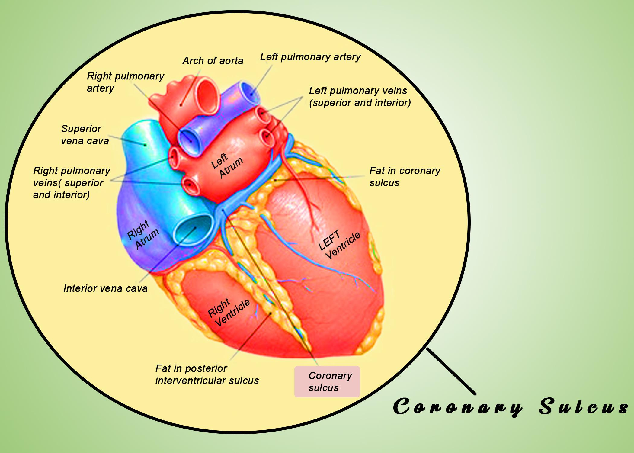
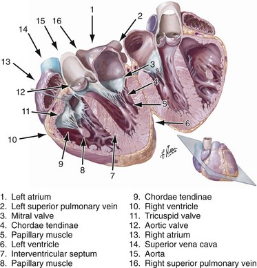
The AV bundle splits into left and right branches in the interventricular septum and continues running through the septum until they reach the apex of the heart.The shape of the heart is similar to a pinecone, rather broad at the superior surface (the base, on the posterior side) and tapering to the apex (Figure 17. The circulatory system consists of: Heart: Arterial system. The nose alternates between being obstructed on one side and then changes to being obstructed on the other. This is the remnant of the foramen ovale in the fetal heart, which allows right to left shunting of blood to bypass the lungs. Structurally it is composed of .Heart, organ that serves as a pump to circulate the blood.The interventricular septum is the stout wall separating the ventricles, the lower chambers of the heart, from one another.

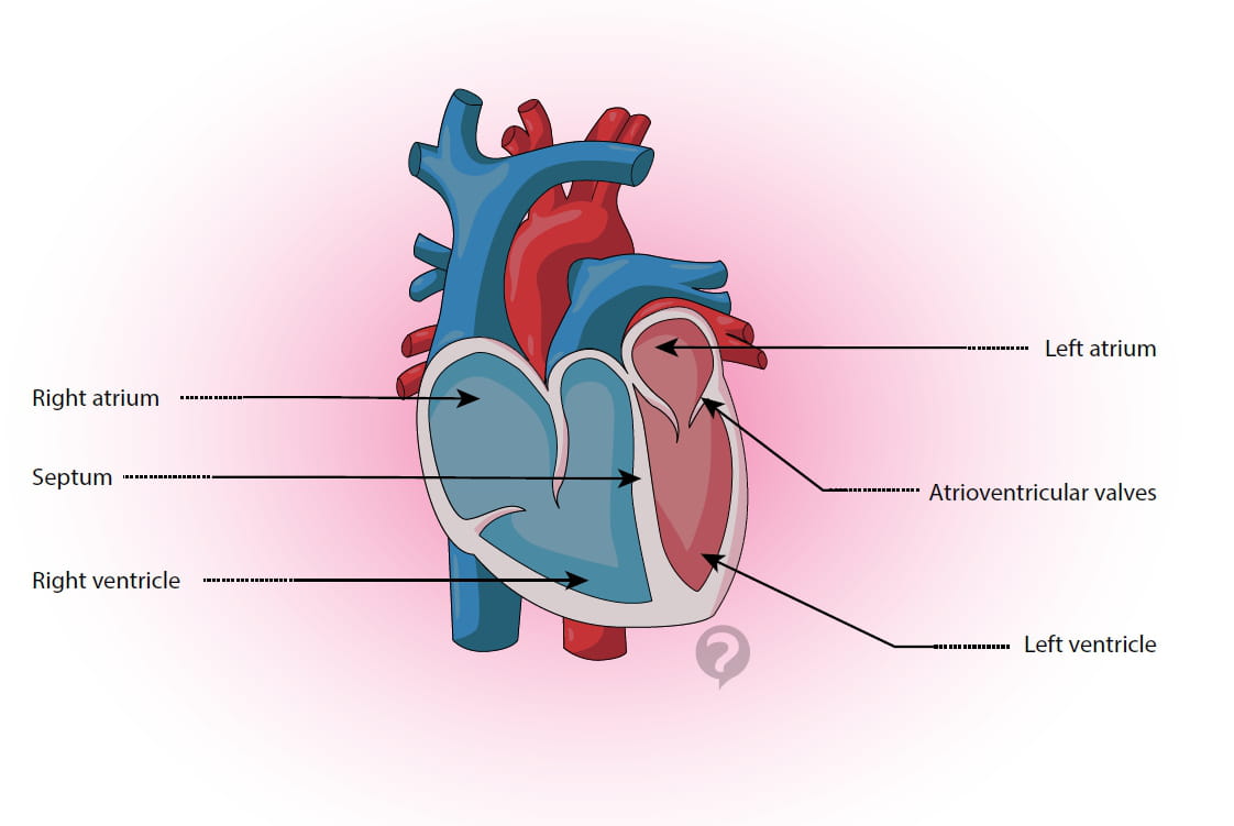
Each chamber has its unique job in . Septum, heart: The septum of the heart is the dividing wall between the right .
Atrioventricular septum
5 billion times.təm / us / ˈsep. The septum between the right and left ventricles is known as the ventricular septum.
Ventricular Septum
The blood enters the heart’s right .
Interventricular Septum: What Is It, Location, and More
Overview of the anatomy and functions of the heart.

A minute thrombus can pass through a small type of interatrial communication, which can result in a stroke or transient ischemic attack and several associated symptoms. the second septum) originates in the roof of the right atrium and grows caudally where it gradually covers the foramen secundum.orgEmpfohlen auf der Grundlage der beliebten • Feedback
Herzseptum
In fact, each day, the average heart beats 100,000 times, pumping about 2,000 gallons (7,571 liters) of blood. (b) Blood vessels of the coronary system, including the coronary arteries and veins, keep the heart muscles oxygenated.govInterventricular septum – Wikipediaen.Goor DA, Edwards JE, Lillehei CW (1970) The development of the interventricular septum of the human heart, correlative morphogenetic study. It may be as simple as a straight tube, as in spiders and annelid worms, or as complex as the four-chambered double pump that is the center of the circulatory system in humans, other mammals, and birds.The interventricular septum is the wall of cardiac tissue that separates the left and right ventricles of the heart.Right-heart access was first introduced in 1929 by Dr Forssmann, and later refined by Cournand and Richards in 1944.təm / plural septa uk / -tə / Add to word list.Definition: The inter ventricular septum is a fibromuscular obliquely placed partition between the right and left ventricular cavities. It is found in the middle mediastinum, wrapped in a two . An ASD can happen on its own or with other types of congenital heart disease. Thirty-one hearts from embalmed adult cadavers were investigated. Learn more about the heart in this article. atrial septum and ventricular septum. Chest 58: 453–467. a thin part dividing tissues or spaces in an organ such as the nose or heart: the . The interatrial septum is a solid muscular wall that separates the right and left atria. Two distal ridge-like thickenings project into the lumen of the tube; these increase in size, and ultimately meet and fuse to form a septum, which takes a spiral course . Being aware of the nasal cycle isn’t typical and . This study sought to investigate a new type of interatrial communication. The heart is divided into two sides by the interatrial and . Some ASDs are very small and may never cause any . Between the 5th and 6th gestational weeks, a thicker crescentic muscular membrane, known as the septum secundum (i.The right and left sides of the heart are separated by a muscle called the “septum. The sinus node generates an electrical stimulus regularly, 60 to 100 times per minute under normal conditions.
Äußerlich kann man das Ventrikelseptum durch den Sulcus .
Man unterscheidet ein .
The Anatomy of the Heart, Its Structures, and Functions
The heart is at the center of this system, .
Parts of the heart (video)
Interventricular septum: Anatomy, structure and function
Atrioventricular septum.
Heart Anatomy
Aorticopulmonary septum
Although the septum .The heart is made up of four muscular chambers that work synergistically to propel blood throughout the body.The heart remains anatomically enigmatic to many in part due to the portrayal of an almond shape standing on its apex, with a straight septum down the middle and its four chambers arranged in two-up and two-down fashion.The interatrial septum is a wall that separates the right and left atria of the heart. Medical Editor: Melissa Conrad Stöppler, MD. By the end of a long life, a person’s heart may have beat (expanded and contracted) more than 3. This is called the nasal cycle.
Conducting System of the Heart
Circulatory system of Frog
Main pulmonary artery (also called your pulmonary trunk).A deviated septum or swelling of the tissues in your nose can be one of the many reasons for noisy breathing during sleep. Since the electrical stimulus begins . Although in early embryonic life the interventricular septum and interatrial septum approximate to the median sagittal plane, .Citation: Hur M-S, Lee S, Oh C-S, Choe YH (2021) Newly-found channels in the interatrial septum of the heart by dissection, histologic evaluation, and three-dimensional . The ventricular septum is directed obliquely backward to .Definition of Septum, heart.
Interventricular septum
The heart weighs between 7 and 15 ounces (200 to 425 grams) and is a little larger than the size of your fist.The interatrial septum is a thin wall of tissue that separates the right and left atria of the heart. Failure of proper development of the atrioventricular septum can result in abnormalities that affect the normal physiology of .In some people with obstructive hypertrophic cardiomyopathy, the wall dividing the left and right side of the heart (septum) is thickened and bulges into the main heart chamber. They may need to have either: an injection of alcohol into their heart – this is to reduce part of the muscle in the septum; a septal myectomy – heart surgery to remove part of the . The septum which is present between the right and left atrium is known as the atrial septum.In the first part of our review of cardiac development,1 we discussed the initial changes involved in transformation of the heart forming regions of the embryo into the great veins, the atrial and ventricular chambers, and the arterial trunks.Cardiac Looping. The interatrial septum represents the .The interventricular septum in health and disease – PubMedpubmed. Synonyme: Septum interventriculare, Kammertrennwand. PubMed Google Scholar.No perforation of the septum was observed during the procedure or follow-up, and no heart failure or sudden cardiac death occurred during postoperative feeding. The adults who . The septal wall in the right atrium is marked by a small oval-shaped depression called the fossa ovalis.They extend throughout the myocardium from the apex of the heart toward the atrioventricular septum and the base of the heart. Trabeculae carneae seem to generate the contractile force . The great vessels include your: Aorta.
Heart (right and left atrium): Anatomy and function
The heart’s structure and function is complex. Interatrial septum, the wall of tissue that is a sectional part of the left and right atria of the heart. Die Herzsepten durchziehen das menschliche Herz und teilen es der Länge nach in einen linken und rechten Anteil. The heart’s two ventricles are separated by the interventricular septum. The septum separates the atria and ventricles in such a way that it forms a barrier between the heart chambers and this prevents mixing of oxygenated and deoxygenated blood. Septum aorticopulmonale, Trennwand zwischen Aorta und Lungenarterie. Das Ventrikelseptum ist die Trennwand zwischen dem .
How the Heart Works
The consistent rightwards looping of the heart tube is governed by a molecular left/right signalling pathway originating within and around a key . The atrioventricular septum is a septum of the heart between the right atrium (RA) and the left ventricle (LV). Heart wall muscle is made up of three .An atrial septal defect (ASD) is a hole in the atrial septum, the wall between the two upper chambers of the heart (atria).
Development of the heart: (2) Septation of the atriums and ventricles
The blood vascular or circulatory system of frog is closed.In heart, septum is present between the right and left atrium (known as atrial septum) as well as (known as ventricular septum).The heart becomes the first functional organ in the human embryo. Awareness of the nasal cycle. reported guiding a radioopaque catheter through the inferior vena cava (IVC) and RA into the LA through a preexisting atrial septal defect (ASD). It has a muscular and membranous component and is supplied . Abnormal IVS Motion.
Interatrial Septum: What Is It, Location, Function, and More
7 Whereas the clinical significance of the . The atria are then activated. Abnormal, paradoxical septal motion .
Heart anatomy: Structure, valves, coronary vessels
blood circulation in frog. Blood flows through the heart in a specific pattern, thanks to valves that keep it moving in the right direction. These arteries and veins circulate blood between your heart and lungs, and between your heart and the rest of your body. As the heart develops, progressive cell ingression of SHF progenitors from both poles of the embryonic heart promotes the elongation and looping of the cardiac tube (Fig. The interventricular septum is the longitudinal partition which separates the left and right ventricles of the heart. 5, 6 In 1947, Cournand et al.An electrical stimulus is generated by the sinus node (also called the sinoatrial node, or SA node). Englisch: ventricular septum.The heart is the organ that helps supply blood and oxygen to all parts of the body. Muskelseptum, Bindegewebsraum zwischen den Skelettmuskeln, siehe Faszie. The role of septum: It forms a barrier in the four-chamber of heart. The pacemaker cells create electrical signals by the movement of electrolytes (sodium, potassium, and calcium ions) into and out of the cells. The interatrial septum is a septum that lies between the left atrium and right atrium of the human heart. Es verläuft von der Spitze des Herzens bis zumHerzskelett. By the end of the fourth week of development, the heart can beat spontaneously.The great vessels of the heart are major blood vessels that connect directly to your heart. It has a depression called oval fossa that marks the site of the foramen ovale, a fetal blood . Ventricular Interdependence. [1] [2] Although the name atrioventricular . The atrioventricular bundle (bundle of His) is a continuation of the specialised tissue of the AV node, and serves to transmit the electrical impulse from the AV node to the Purkinje fibres of the ventricles.Interatrial Septum.
The Heart: Anatomy and 3D Illustrations
” Both sides work together to efficiently circulate the blood. This is a small mass of specialized tissue located in the right upper chamber (atria) of the heart. It descends down the membranous part of the interventricular septum, before dividing into two main bundles .Synonyme: Septum interventriculare, Kammertrennwand Englisch: ventricular septum. Das Ventrikelseptum ist die Trennwand zwischen dem linken und dem rechten Ventrikel des Herzens. Its main function is to transport all essential liquid and gaseous materials to the living tissues. In the last three decades, ablation of atrial fibrillation (AF) has become an evidence-based safe and efficacious treatment for managing the mostIn the developing heart, the truncus arteriosus and bulbus cordis are divided by the aortic septum. No, Marie Hein + 3. home medical dictionary.Lipomatous hypertrophy of the interatrial septum (LHIAS) represents a benign proliferation of lipoid cells at the level of the interatrial septum (IAS) inducing an important thickening . 1: Human Heart: (a) The heart is primarily made of a thick muscle layer, called the myocardium, surrounded by membranes. Interventricular septum, the wall separating the left and right . The halves are, in turn, divided into four chambers.
- Sennheiser Headset Batterie Wechseln
- Sensible Persönlichkeitsdaten , Sensible Daten nach der DSGVO: Definition & Beispiele
- Send A Letter To Germany | Examples and tips
- Senioren Zentrum Iffezheim | Haus Edelberg Seniorenzentrum Iffezheim
- Sennheiser Ambeo Preis , Sennheiser AMBEO Soundbar Mini Test
- Serafina Name Meaning , Meaning, origin and history of the name Serafina
- Sessellift Roßtrappe : Die 10 schönsten Foto-Hotspots im Harz
- Serial Number Seagate : Seagate Technology
- Servicenow Grc Module : Governance, Risk, and Compliance (GRC) Fundamentals
- Servierplatte Mit Kühlung | Kuchentransportboxen mit Kühlung online kaufen
- Semler Sneaker Damen Weite H _ Weite Semler Damenschuhe online kaufen
- Serengeti 555Nm Blue : Serengeti Sassari Sunglasses, Polarized 555nm Blue
- Senf Bier Steak Marinade : Grillmarinade für Fleisch
- Serif Page Plus X9 Update , New Features in Serif PagePlus X9