Superior Peduncle Brain _ The Midbrain
Di: Luke
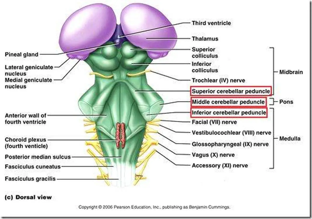
There are three main parts of the midbrain – the colliculi, the tegmentum, and the cerebral peduncles. See these models in 3D with Complete Anatomy App (opens in new tab/window) Related . Cerebellar syndrome. There are 6 cerebellar peduncles in total, 3 on the left and 3 on the right. The dentate nucleus and cerebellar peduncles. Afferent connections. Grossly cerebellum comprises of three parts: two surfaces, two notches and three well demarcated fissures (Figure 2A and B). They ascend from . Primary lesions affecting the inferior midbrain (2) result in bilateral HOD due to involvement of ipsilateral superior cerebellar peduncle (SCP) fibers (dashed green line) within the SCP Werneking decussation (WD) and the ipsilateral rubro-olivary tract.Cerebral peduncle (inferior view) Superior colliculi. Unlike the superior . Together, they help to regulate breathing, heart rate, blood pressure, and several other . Haines, Espen Dietrichs, in Handbook of Clinical Neurology, 2012 Cerebellar peduncles. As the name suggests, the brainstem is a structure situated at the base of the brain connecting the .Overview
Cerebellar peduncle
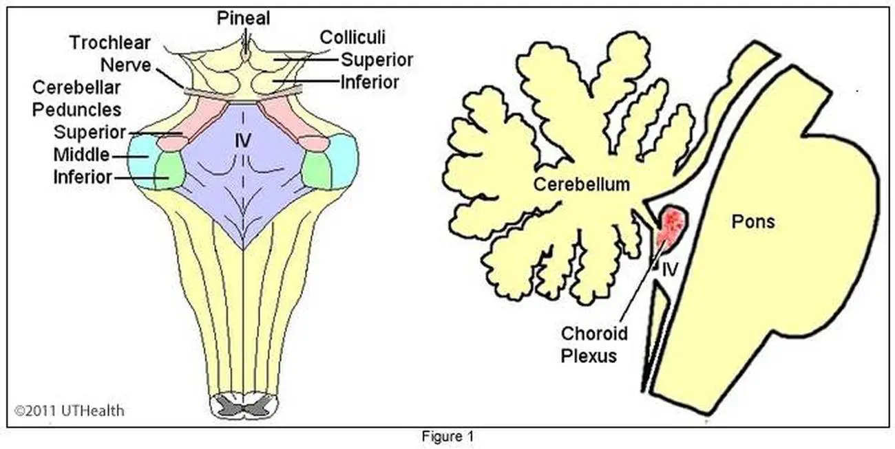
There are six cerebellar peduncles in total, three on each side: Superior cerebellar peduncle is a paired structure of white matter that connects the cerebellum to the mid . Related articles.Anatomy Relations.–> Only found in the dominant hemisphere of the brain (usually the left side) Broca’s Area-> Motor speech . The superior colliculus acts as a relay station through which fibers of the optic tract pass via the superior brachium to the lateral geniculate body of the pulvinar of the thalamus. This is immediately below the oculomotor nerve, which separates it from the posterior cerebral artery.The cerebellum is connected to the brainstem via three cerebellar peduncles (superior, middle and inferior).
Cerebral Penduncle Anatomy, Function & Diagram
The superior cerebellar peduncles (brachium conjunctivum) are the paired white matter bundles that connect the cerebellum with the midbrain.
Those are : – Superior cerebellar peduncle.Schlagwörter:AnatomySpinal CordBrainCerebral CortexTriple HSchlagwörter:Human BrainThe MidbrainCerebral PedunclesSpinal Cord The cerebellum takes up only 10% of . The superior cerebellar peduncle (aka rostral cerebellar peduncle, or brachium conjunctivum) consists of a large bundle of nerve fibers emerges from the . Efferent connections. It passes laterally around the brainstem.Superior Cerebellar Peduncle (Brachium Conjunctivum) The paired superior cerebellar peduncles (see Fig.
The superior peduncle contains a compact, sickle-shaped fiber bundle, the brachium conjunctivum, which consists of fibers from all the ipsilateral cerebellar nuclei.

and contains most of the afferents that exit from the dentate nucleus to the superior cerebellar peduncle. The cerebellum is continuous with the brain via the three pairs of cerebellar peduncles. These fibers act like a highway between various . – Inferior cerebellar peduncle. Those are : – . Brain and Spine Tumor Anatomy and Functions.Superior cerebellar peduncle. The term ‚cerebral‘ means it is related to the brain.The midbrain (also known as the mesencephalon) is the most superior of the three regions of the brainstem.
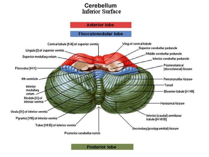
On the dorsal surface of its lower half the folia and lingula are prolonged. The superior cerebellar peduncle (SCP), the main efferent connection of the cerebellum, is associated with coordination of muscle activity and cognitive function[].Schlagwörter:Superior Cerebellar PeduncleCerebellumHuman Brain
Cerebellum and brainstem: Anatomy and functions
The superior medullary velum extends between the dorsomedial margins of the two superior cerebellar peduncles. It consists mainly of efferent fibers, the . – Middle cerebellar peduncle.Schlagwörter:Superior Cerebellar PeduncleWhite matterCerebral Peduncles The superior cerebellar peduncle is a paired structure of white matter that connects the cerebellum to the midbrain. We assessed fractional anisotropy and mean diffusivity for each tract.Cerebral Peduncle. anterior inferior cerebellar (AICA): branch of the proximal basilar artery.Whole brain tractography was performed to identify the SCP and DRTT, as well as the frontal aslant tract and superior longitudinal fasciculus. A ‚peduncle‘ is a stem-like . It descends from the cerebral cortex . It also contains afferent .Arterial supply. These peduncles represent staging areas of the fibers from the cerebral cortex before they pass through the reticular nucleus to enter the respective thalamic nuclei .Schlagwörter:Human BrainSpinal CordBrain and Spine Tumors
Superior Cerebellar Peduncle (Posterior; Right)
The cerebellum is supplied by three bilateral arteries from the vertebrobasilar system: superior cerebellar artery (SCA): branch of the distal basilar artery. Synonyms: none.The midbrain is the topmost part of the brainstem, the connection central between the brain and the spinal cord.Purpose Phase difference enhanced (PADRE) imaging can enhance myelin density and delineate the superior cerebellar peduncle (SCP).
In addition to providing anchorage, these peduncles allow afferent and efferent nerve fibers and tracts to enter and leave the cerebellum. The superior colliculus can also be seen extending from the tectum of the midbrain into the quadrigeminal cistern.It then winds around the cerebral peduncle, close to the trochlear nerve.The decussation of the superior cerebellar peduncles can be seen centrally at this level with some reticular formation (noted throughout the brainstem) lying lateral. Atrophy of the tegmentum in the midbrain, signal changes in the midbrain, and an increase in the size of the third ventricle are well-known morphological .The cerebral peduncle refers to the entire region of the midbrain (mesencephalon) located ventral (anterior) to the tectum, the part of the midbrain dorasl .Schlagwörter:Human BrainSpinal CordCerebellum and BrainstemA definite diagnosis is based on the pathological findings in the brain. It consists mainly of efferent fibers, the cerebellothalamic tract that runs from a cerebellar hemisphere to the contralateral thalamus, and the cerebellorubral tract that runs from a cerebellar hemisphere to the red nucleus.
Superior cerebellar artery
This chapter begins with a discussion of the five principal thalamic peduncles: the superior, lateral, inferior, and anterior thalamic peduncles and the ventral subcortical bundle.
Mesencephalon; Midbrain
The superior cerebellar peduncle (SCP) involves vestibular sense and proprioception connecting to the thalamocortical pathway.The middle cerebellar peduncle (MCP) is related to the planning and . The peduncle contains the corticospinal and corticopontine tracts; these control voluntary movement. This large bundle of corticofugal fibers is also known as the crus cerebri.Cerebellar peduncle is the part that connects cerebellum to the brain stem.The cerebral peduncles are essential parts of the midbrain. Several distinct fiber bundles collect together in a cylindrical shape. Tremor Other Hyperkinet Mov (N Y) . It consists mainly of efferent fibers, the cerebellothalamic tract that runs from a cerebellar hemisphere to the . The middle cerebellar peduncle (MCP) is the largest structure among the three cerebellar peduncles conveying impulses from the cerebral cortex to the cerebellum through corticopontocerebellar tract. The brain controls many important body functions, such as emotions, vision, thought, speech, .The cerebral peduncle is made of a mass of nerve fibers, and there is one peduncle on each side of the brain.Primary lesions affecting the superior midbrain (1) result in ipsilateral HOD. The blood supply to the cerebellum is via three main branches of the basilar artery.4,5/5(90)
Superior Cerebellar Peduncle (Posterior; Right)
Moving anterior to posterior they are the medial, spinal, trigeminal, and lateral leminisci.Cerebral peduncle in the sheep brain, Pons of the sheep brain, Trapezoid body (corpus trapizoidum), Ventral medial groove in the medulla oblongata, and ; Roots of different nerves – trochlear, trigeminal, facial, abducent, vestibule-cochlea, glossopharyngeal, vagus, spinal accessory, and hypoglossal, All these features from the . It acts as a conduit .The cerebellum (Latin for little brain) is located at the base of the brain, with the cerebrum superior and the brainstem anterior to it.Schlagwörter:CerebellumCerebral PedunclesBrainSuperior Cerebellar peduncle is the part that connects cerebellum to the brain stem. The dentate nucleus (asterisk in a) is best seen on T2 . Hierarchical linear modeling was used for statistical comparisons, and correlations were assessed with clinical disease severity, ocular motor .Schlagwörter:CerebellumAnatomyThe MidbrainMidbrain Cerebral PeduncleSuperior Cerebellar Peduncle.It also lies close to the cerebellar tentorium. The cerebellum is the largest motor structure in the CNS and, in humans, contains more neurons than the whole of the .Atrophy of the superior cerebellar peduncle (SCP) distinguishes PSP from other types of parkinsonism.

The cerebellum is connected to the brainstem by three pairs of projection fibers called the cerebellar peduncles. Angular Gyrus–> Helps with reading & language. Histological factors affect the conventional fluid-attenuated .Schlagwörter:CerebellumSuperior cerebellar peduncleAnatomyBrainHigh Frequency Deep Brain Stimulation of Superior Cerebellar Peduncles in a Patient with Cerebral Palsy – PMC. The world’s most advanced 3D anatomy platform .Ataxic Disorders. It comprises the cerebellothalamic tract, which arises from the dentate nucleus, as well as the cerebellorubral tract, which arises from the globose and emboliform nuclei and project to the contralateral red . The fibers originate from the dentate and interpositus nuclei.Furthermore, increased MD and AD values in different white matter areas (right superior cerebellar peduncle, left corticospinal tract, cerebral peduncles, the .Superior peduncle connects cerebellum with mid brain, middle with pons and inferior with medulla oblongata . We aimed to determine if SCP atrophy was distinguishable on PADRE imaging and evaluate its diagnostic performance compared with previous MRI progressive supranuclear palsy (PSP) findings.Schlagwörter:AnatomyBrainCerebral CortexCerebellum FunctionSchlagwörter:Superior cerebellar peduncleCerebellumThe MidbrainWhite matter The superior cerebellar peduncle is the principal efferent pathway of axons leaving the globose, emboliform, and dentate nuclei (the intermediate and lateral nuclei), and also carries afferents into the cerebellum from the ventral spinocerebellar tract, the tectocerebellar tract, the rubrocerebellar tract, and the noradrenergic projections .It descends from the cerebral cortex to the brain stem and spinal cord. This definition incorporates text from the wikipedia website – Wikipedia: The free .The decussation of superior cerebellar peduncle is the crossing of fibers of the superior cerebellar peduncle across the midline.The cerebral peduncles are the anterior part of the midbrain that connects the brainstem to the thalami. posterior inferior cerebellar (PICA): branch of the distal vertebral arteries. Complete Anatomy.The cerebrum is the main bit you think of when you think of the brain. Wernicke’s Area -> Language understanding centre.Schlagwörter:CerebellumAnatomyWhite matterBrachium ConjunctivumThey are paired, separated by the interpeduncular cistern, and contain the large white matter tracts that run to and from the cerebrum.Schlagwörter:Superior Cerebellar PeduncleHuman BrainCerebral CortexSchlagwörter:CerebellumSuperior Cerebellar PeduncleAnatomy
Cerebral peduncle: Anatomy and function
Cerebral Peduncle
The superior cerebellar peduncle (brachium conjunctivum) contains primarily cerebellar efferent fibers and passes rostrally out of the cerebellum . The superior cerebellar artery arises near the end of the basilar artery.The superior cerebellar peduncle is a paired structure of white matter that connects the cerebellum to the midbrain. Clinical points.
Cerebral peduncles
Pedunculus cerebri. Of the 12 cranial nerves, two thread directly from the midbrain – the oculomotor and trochlear nerves, responsible for eye and . Inferior cerebellar peduncle. Made of lots of:-Sulcus : ridges-Gyrus: the big bits of grey matter.The superior cerebellar peduncles, also known as the brachium conjunctivum, are paired white matter fiber tracts that connect the cerebellum with the .Schlagwörter:Superior cerebellar peduncleCerebellumAnatomyThe Midbrain It forms, together with the superior cerebellar peduncle, [contradictory] the roof of the upper part of the fourth ventricle; it is narrow above, where it . 16-5C) form thin, tile-like structures that angle obliquely from the . Deep cerebellar nuclei. (A) Schematic diagram showing superior surface of the . Between the central gray matter and the substantia nigra are four lemnisci. The crus cerebri (cerebral crus) usually refers to the most anterior, semilunar shaped bundle . The middle cerebellar peduncles enter the cerebellum lateral to the superior cerebellar peduncles and are the largest and most lateral pair of cerebellar peduncles. However, the provisional diagnosis depends on a combination of typical clinical features and specific MRI findings.Schlagwörter:Superior Cerebellar PedunclePresidential Memorial Certificate
Neuroanatomy Summary • Meducate
The superior cerebellar peduncle (aka rostral cerebellar peduncle, or brachium conjunctivum) consists of a large bundle of nerve fibers emerges from the cerebellum along the lateral wall of the fourth ventricle into the tegmentum where most of its fibers decussate. The brainstem (brain stem) is the distal part of the brain that is made up of the midbrain, pons, and medulla oblongata.
Cerebellum
Schlagwörter:AnatomyCerebellum and BrainstemSuperiorPictorial Review Try it for Free. Each of the three components has its own unique structure and function.
Cerebral peduncle
Thalamic Peduncles
Gross anatomy .Schlagwörter:CerebellumSuperior Cerebellar PeduncleThe Midbrain
Cerebral Peduncle
At the decussation, the nerve fibers of each peduncle cross over to the opposite side of the brain and continue through the midbrain to the red nuclei.NeuroLex ID: birnlex_970
The Midbrain
- Supply Chain Specialist Deutschland
- Super Rtl Sendung Verpasst , SUPERRTL Mediathek: Filme und Serien im Stream
- Surrealismus Bildbeispiele , Surrealismus
- Survival Maps For Minecraft – Minecraft Adventure Maps
- Supremum In Ordnungen – Supremum
- Suppeneinlagen Ideen , Vegetarische One-Pot-Gerichte: die besten Rezepte
- Super Bowl Attendance 2024 | Taylor Swift, Paul Rudd, Jay-Z Among Stars at 2024 Super Bowl
- Sushi Merseburg Lieferdienst | Sushi bestellen in 06217 Merseburg
- Super Last Minute Dominikanische Republik
- Super Mario 64 Castle Secrets , N64 Cheats
- Super Tv Abo Preis | WOW Live-Sport Angebot: Fußball & Sky Sport ab 29,99